
A woman who can't talk but can sing

A 30-year-old woman who we’ll call Issy was brought to the Emergency Department due to one-sided facial paralysis and weakness in her right arm and leg.
Every morning, Issy performs her morning ritual before going to work. That morning was no different. Issy woke up at about 6:30 AM to the sound of her favorite song sung by her husband.
Issy and her husband had a ritual of singing the same song every morning to boost the start of their day.
Issy then made a cup of coffee, took a shower, and headed to her garden before going to work. Once she left the garden, she suddenly collapsed to the floor due to a sudden weakness in her right leg. Her husband worked in construction, so he left before her. Several hours passed, and her neighbor found Issy lying on the garden floor, struggling to get up. When asked how it happened, she couldn't give him a proper answer since she had slurred speech and was extremely confused.
In a panic, her neighbor called an ambulance, which got her to the Emergency Department.
Issy arrived at the Emergency Department at 11:00 AM, and the doctor immediately suspected that she was having a stroke and ordered a CT scan.
A standard stroke protocol starts with a non-contrast head CT, which may identify the early signs of stroke, but more importantly, can exclude intracerebral hemorrhage (meaning bleeding in the brain) and other processes in the brain that might resemble an acute ischemic stroke, such as brain tumors.
Non-contrast CT is useful in the evaluation of acute intracranial hemorrhage as it produces good contrast between the high attenuating (“bright”) clot and the low attenuating (“dark”) cerebrospinal fluid.
On a non-contrast CT scan, stroke usually presents as a subtle blurring and decreased attenuation of the grey-white junction due to edema (meaning swelling of the brain tissue between the white and gray matter of the brain).
This tool’s availability and speed make it very useful in initially evaluating suspected stroke patients.
The next step in diagnosis is either a CT or MR angiogram of the head and neck with perfusion that images from the aorta up to the brain.
The angiogram aims to assess for areas of stenosis or occlusions that may explain the symptoms. The purpose of the perfusion scan is to determine the extent of tissue that has already been damaged versus the extent that is at risk of damage, or in medical terms, the core versus the penumbra.
This determines whether or not the patient is a candidate for mechanical thrombectomy (remember this).
If the patient gets a CT and CT angiogram, they will eventually need an MRI (magnetic resonance imaging) as well.
There are also several important laboratory studies to be performed. One is a coagulation panel, particularly in the setting of a patient on warfarin, an anticoagulant medication that prevents blood from forming a thrombus that can occlude major blood vessels and lead to ischemic stroke. Blood glucose levels should be obtained in addition to a complete blood count and basic metabolic panels. Blood glucose levels are especially important since hypoglycemia (low blood glucose levels) can mimic stroke and can be life-threatening if not recognized and treated promptly.
Lastly, cardiac enzymes should be done as well to assess for cardiac abnormalities since a thrombus can be formed inside one of the heart chambers in some cases of irregular heart rhythms such as atrial fibrillation.
After a stroke has been identified, the patient typically will need to get an EKG and a transthoracic echocardiogram (meaning a heart ultrasound through a patient's chest wall) to determine whether there is an intracardiac thrombus or cardiac abnormality that may be the source of the patient's stroke.
Generally, when we talk about stroke, we must differentiate between the 3 main types.
The first type is the ischemic stroke. Ischemic from Ancient Greek ἴσχαιμος (“stoppage of blood flow”) refers to a stroke that happened due to a blockage of one or several blood vessels that perfuse the brain. Since the brain is the most energy-demanding organ in our body, without oxygen and other nutrients, the brain cells quickly die off. Unfortunately, nerve cells are one of the few types of cells that can't regenerate themselves.
The second type of stroke is hemorrhagic stroke, meaning the stroke is due to a rupture of certain blood vessels that perfuse the brain. In a hemorrhagic stroke, there are two main problems: the first problem is that the damaged blood vessel cannot adequately transport nutrients to the proper part of the brain. The second problem is that the accumulation of blood inside the brain pushes the brain tissue, damaging it consequently.
The third part of a stroke is a so-called transient ischemic attack, or TIA for short. TIA is a stroke that lasts only a few minutes. It occurs when the blood supply to part of the brain is briefly or temporarily interrupted, which leads to neurological symptoms similar to a real stroke. Even though TIA is not considered as dangerous as a real stroke, it is a good predictor of possible strokes in the future.
While waiting for a CT scan, the doctor performed a neurologic exam.
The most important part of the neurological exam is to perform the National Institute of Health Stroke Scale or NIHSS for short.
The components that are tested in NIHSS include sensation, strength, and coordination in all 4 extremities; production and comprehension of speech, including naming and repetition; visual fields; orientation to self and time; and symmetry and sensation of the face.
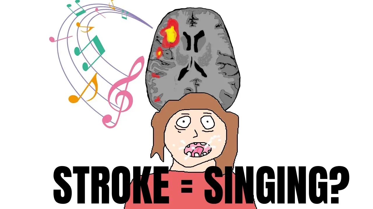
In the case of Issy, she had a large middle cerebral artery stroke (MCA), which is one of the most common and easiest-to-recognize types of stroke. It tends to present with the major deficits that one thinks of when thinking of a stroke, such as unilateral flaccidity, forced gaze deviation, visual field cuts, and, if in the dominant hemisphere, speech deficits.
In Issy, unilateral weakness, facial droop, and speech deficits were present.
In the acute setting, the treatment of MCA stroke includes two interventions.
The first is the IV tissue plasminogen activator (TPA), and the second is thrombectomy, as we previously mentioned. IV TPA is a type of medication that works by dissolving blood clots that block blood flow to the brain. A prerequisite for the administration of IV TPA is that symptoms of a stroke have to occur within the timeframe of 4.5 hours.
Since more than five hours passed between Issy's collapse and her arrival at the ER, our patient had to go through another procedure called mechanical thrombectomy.
Mechanical thrombectomy is an endovascular technique for removing blood clots from the brain after an ischemic stroke by inserting a thin tube that reaches the occluded blood vessel.
For thrombectomy, the symptoms mustn’t last for more than 24 hours before intervention.
In addition, it is necessary to check vital signs, particularly the patient's blood pressure as hypertension and hypotension can both be associated with neurological symptoms, and blood pressure must be under 180/110 before administration of IV TPA since they can cause unwanted bleeding.
Issy underwent a mechanical thrombectomy and was hospitalized for 2 weeks. Luckily for her, she had an almost complete recovery, but with prominent expressive aphasia that sadly didn't go away even with regular sessions with a speech therapist.
In other words, Issy lost her voice completely.
Even though her husband was her constant support through her recovery, he couldn't fail to notice that the loss of his wife's voice brought her a sense of alienation from her husband and their merry mornings singing their favorite song.
But what happened next was unexpected, to say the least.
One morning, Issy woke up and continued through her daily routine. Usually discouraged when she remembers that stroke left her without a voice, this morning her mind was occupied with something else. Forgetting about her lost voice, she accidentally started singing with almost perfect clarity. She was appalled, to say the least.
But how could this happen?
Let me explain.
Our patient suffered from an ischemic stroke caused by an occlusion of her left middle cerebral artery, which is one of the three major arteries that supply the brain. MCA, among other parts of the brain, supplies the anterior speech area called Broca’s area in the frontal lobe of the brain. This speech area of the brain is located on the left side of the brain in most people; thus, left MCA occlusion leads to damage to this area.
Damage to Broca’s area leads to so-called expressive aphasia, which means an inability to produce speech in your brain and consequently a stuttering speech pattern.
Another type of aphasia is receptive aphasia, in which people can talk fluently but without understanding speech.
A few studies noticed that in healthy people, the right side of the brain lights up on MRI as they are singing. This is not to say that some unknown area on the right side of the brain is solely responsible for the act of singing, because singing combines both music and words, which are mostly produced in the left hemisphere of the brain.
In this case, the left side of the brain, responsible for word recognition, remained unchanged, and when coupled with the right brain area responsible for sound and music, Issy managed to sing even though she lost the ability to speak.