
Death by Rectal Exam: One of the most interesting cases I've ever read

A 66-year-old male was brought to the Emergency Department with a complaint of abdominal distention, meaning he had a swollen abdomen. His other symptoms were insignificant except a decreased appetite, which wasn’t unusual since his last bowel movement was about a week ago. Even though the patient had a past medical history notable for several psychiatric and non-psychiatric comorbidities, none of those could explain the sudden stop of bowel movements. The patient was on a long-term antipsychotic therapy regimen but had no previous gastrointestinal side effects.
The patient's antipsychotic medications included both fluphenazine and lithium, medications that can cause bradycardia (slowing of the heart rhythm) and other changes in the patient's electrocardiogram.
This information will be crucial later in the diagnosis.
On initial presentation to the Emergency Department, the patient was tachycardic and hypertensive, meaning his heart rate was over 100 beats per minute and his blood pressure was over 130/80. The patient was also tachypneic, meaning his respiratory rate (RR) was over 20 per minute. A pulse oximetry test showed 93% oxygen saturation in room air. Pulse oximetry is a test used to measure the oxygen saturation of the blood. It is a non-invasive, painless measure of how well oxygen is being sent to parts of your body furthest from your heart, such as the arms and legs.
On examination of the patient, his oral mucosa was noticeably dry, a usual sign of dehydration. His cardiac exam was unremarkable without murmurs, rubs, or gallops, and his lungs were clear to auscultation bilaterally, meaning both sides. The patient's abdomen was firm and grossly distended and was noted to resemble a full-term pregnant patient. On inspection, dilated superficial veins were noted on his abdomen, which could be explained by the venous blood stasis as a consequence of his dilated abdomen. On palpation, his abdomen was mildly tender but without rebound or guarding, both of which are signs of acute abdominal inflammation.
The patient was awake, alert, and oriented to time, date, and environment.
His lab work showed hyponatremia, meaning a low concentration of sodium in the blood, and hyperkalemia, meaning a high concentration of potassium in the blood. Sodium and potassium are one of the most important electrolytes that serve as messengers to our cells and are especially important to our cardiomyocytes (cells that make up our hearts).
The patient was also anemic with a slightly lower concentration of hemoglobin, a protein found in the blood that carries oxygen throughout our body.
An electrocardiogram was obtained during the Emergency Department evaluation, which showed a sinus tachycardia of 105 beats per minute and peaked T waves in the precordial leads.
Sinus tachycardia refers to a normal heart rhythm with higher than normal beats per minute, and peaked T waves can be an early sign of a heart attack.
In this case, a heart attack can be ruled out since the patient's antipsychotic medication can cause peaked T waves to appear on an electrocardiogram.
The next step in diagnosis was a computerized tomography (CT) scan of the abdomen and pelvis, which showed large dilated loops of colon noted to be up to 12 cm, and the acute care surgical service was consulted for further management. Since the CT showed large dilated loops of the colon, meaning there was an obstruction that prevented stool from going out of the colon, the first step in the management of such a patient includes a digital rectal exam and possible disimpaction.
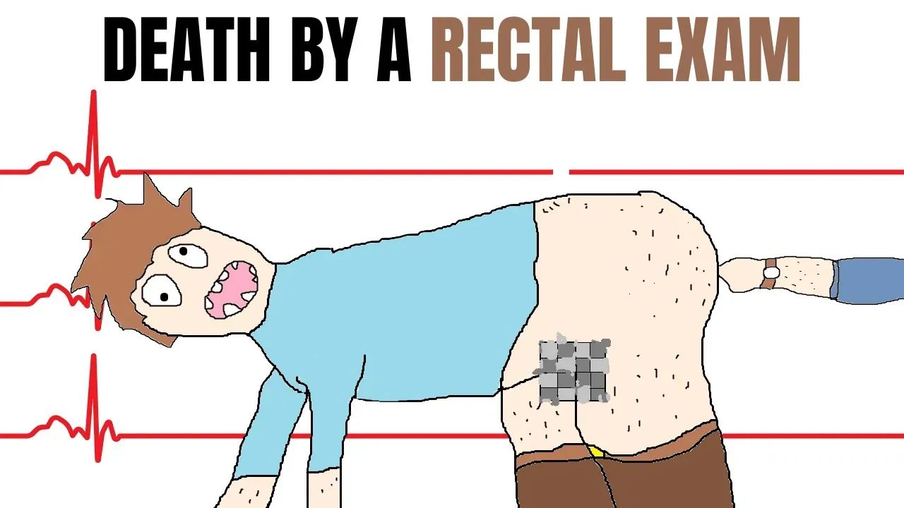
A digital rectal exam is a medical test in which a doctor inserts a gloved, lubricated finger into your rectum and feels the back wall of the prostate gland for enlargement, tenderness, lumps, or hard spots. Even though digital rectal exams can be performed for a variety of reasons, the most common indications are diseases of the prostate, such as prostate cancer or prostate enlargement in older men. Although a digital rectal exam is usually without complications, in some rare instances, it can lead to serious problems, as you are about to read now.
The patient was positioned on his left side, and a soft stool was found on the rectal exam. During the rectal examination, the nurse in the room noted that the patient was apneic, meaning he wasn’t breathing, and a code was called. A code in the medical dictionary refers to a cardiopulmonary arrest that needs immediate start of resuscitative efforts. The patient was unresponsive for less than one minute before the nurse noticed his status change.
Unfortunately, the patient was not on a cardiac monitor at this time.
Since the patient was not on a cardiac monitor at the time, one was placed immediately, and a bradycardic rate was noted. Once again, bradycardic means low heartbeats per minute.
The resuscitation team checked his pulse and found that the patient was in a so-called pulseless electrical activity.
Pulseless electrical activity refers to a condition where your heart stops because the electrical activity in your heart is too weak to make your heartbeat. Pulseless electrical activity is considered an emergency since the heart is no longer able to pump blood effectively. The lack of oxygen that is normally carried through your blood leads to a certain cell death across all organ systems in your body.
Cardiopulmonary resuscitation or CPR was started, and advanced cardiac life support (ACLS) protocol was followed, including intubation. Following multiple rounds of CPR and ACLS medications, spontaneous circulation was obtained. The patient then again went into pulseless electrical activity with a bradycardic rhythm, but ROSC (return of spontaneous circulation) was obtained and remained.
Unfortunately, the patient’s arterial blood gas showed severe acidosis, and the patient was then admitted to the Intensive Care Unit (ICU) on epinephrine, norepinephrine, and vasopressin infusions.
Even though the patient had a normal heart rhythm and a rate of 98 beats per minute, eight days later the patient was confirmed to be dead because of an anoxic brain injury that developed during his arrest, meaning his brain didn’t manage to get enough oxygen, and before-mentioned cell death occurred.
But why did the patient go into cardiac arrest in the first place?
Let me explain.
The human nervous system is divided into the central and peripheral nervous systems. A component of the peripheral nervous system is called the autonomic nervous system. The autonomic nervous system serves as an “involuntary machine” that guides various aspects of our physiology.
It is comprised of the sympathetic and parasympathetic nervous systems. The sympathetic nervous system is responsible for the fight-or-flight response, a series of autonomic reactions caused by a stressful event. In a fight or flight response, your body prepares to face a certain threat by increasing your blood pressure, heart rate, and breathing rate.
On the opposite side, we have a parasympathetic nervous system, which is responsible for lowering your blood pressure, heart rate, and breathing rate and promoting digestion in a state of relaxation. It serves to relax your body (if you will). The biggest parasympathetic nerve in our nervous system is the so-called Vagus nerve, which travels from your brain down to your colon. Even though the vagal nerve doesn’t reach your rectum directly, other parasympathetic nerves are responsible for rectal innervation.
These nerves go both ways. The nerves from the brain send signals to your intestines for them to increase their motility during digestion. In return, the nerve signals are sent to your brain that the passage of stool is happening. This way, your parasympathetic nervous system is activated, and other physiological processes take place since you are no longer in immediate danger.
By performing a digital rectal exam, you stimulate those nerve endings, which activate your parasympathetic nervous system, slowing your heart rate and blood pressure. Normally, these changes are not a cause for an alarm (concern) since they are part of a normal physiological process.
However, this patient already had an electrically unstable heart because of low sodium and high potassium in his blood.
Another risk factor was his antipsychotic regimen, including fluphenazine and lithium, which are both described to cause changes in heart activity, mainly slowing the heart rate.
Usually, a safe digital rectal exam was probably a tipping point that snowballed the patient into cardiac arrest and, unfortunately, anoxic brain injury.